- December 14, 2020
- By Rebecca Copeland
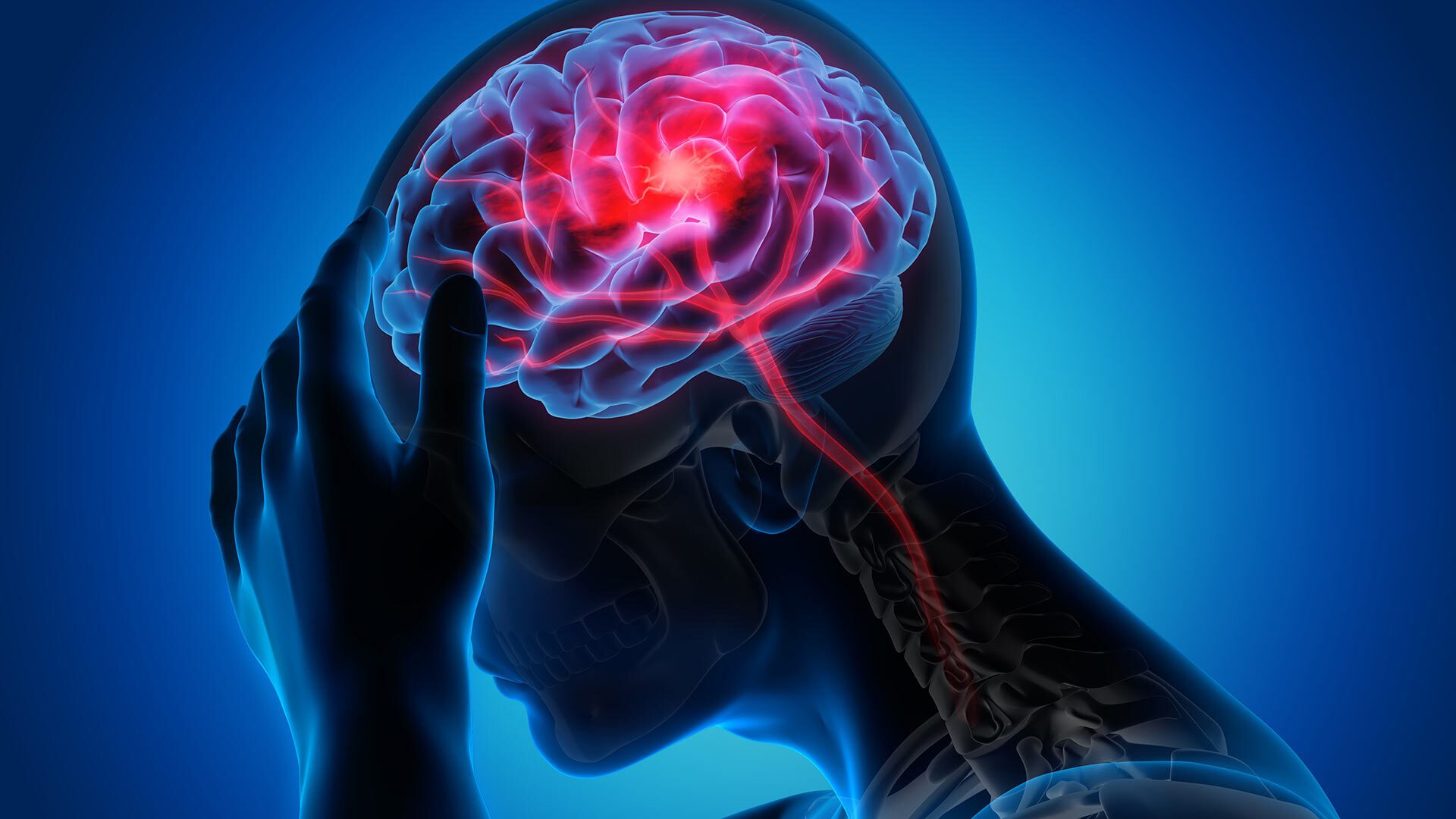
After Julia had a minor stroke, she was thankful for receiving rapid treatment and recovering well. But she soon noticed unexpected aftereffects: In meetings at work, she was unable to participate in the normal back-and-forth, and at home, she couldn’t easily carry on a conversation with her husband while, say, cooking dinner.
When she mentioned her symptoms at her next doctor visit, she found out she was not alone. Julia is experiencing poststroke acute dysexecutive syndrome (PSADES), a common cognitive dysfunction after even minor strokes. Although stroke patients have long reported these cognitive difficulties, evidence has mostly been anecdotal until now.
A new study by University of Maryland, Johns Hopkins University and New York University researchers for the first time provides measurable physical evidence of diminished neural processing within the brain after a stroke, and suggests that PSADES is the result of a global connectivity dysfunction.
The research published today in the Proceedings of the National Academy of Sciences of the United States of America (PNAS) by Professor Jonathan Simon—with appointments in electrical and computer engineering, biology and the Institute for Systems Research—his former postdoctoral researcher Christian Brodbeck, now a visiting assistant professor at the University of Connecticut, and UMD Ph.D. student Joshua Kulasingham.
The study’s lead author was Johns Hopkins School of Medicine’s Associate Professor Elisabeth Marsh; other collaborators included Professors Rafael Llinas and Dania Mallick, her colleagues in the Department of Neurology, and NYU Grossman School of Medicine Research Professor Rodolfo Llinas.
“We tend to think that certain parts of the brain are responsible for specific functions, but in reality you need your entire brain to think clearly and complete tasks,” Marsh said. “In this study we show how a small lesion anywhere can disrupt the cognitive network and result in a global dysfunction.”
The researchers used magnetoencephalography (MEG) to look at the brain functioning of patients who recently experienced minor strokes. MEG is a non-invasive neuroimaging technology that employs very sensitive magnetometer sensors to make high-speed recordings of naturally occurring magnetic fields produced by electrical currents inside the brain.
Patients had their magnetic fields recorded as they completed word and picture matching tasks. These tasks all involved memory, memory search or identification. In some tasks the patient needed to speak the answer, while others required them to press a “yes” or “no” button. An age-matched control group of people who had not suffered a stroke also completed the tasks and were recorded.
When the two sets of MEG recordings were compared, stroke patients’ recordings exhibited distinct characteristics that were different from the control group. For example, the signals within their brains were noticeably more subdued, appearing more like rolling hills rather than mountain peaks. This is an indicator that the brain is processing less efficiently. The stroke patients also took about twice as long as the control group to complete the tasks.
These patterns of inefficient processing suggest a dysfunction in the brain’s distributed network—a disruption of the network’s dynamics.
“If the problem is not because of the lesion itself, affecting visual processing regardless of where the lesion is located, and you can’t see what is wrong in an MRI or CAT scan, then we conclude that the issue is more global: how the brain talks to itself,” said Simon. “This shows that even a minor stroke, whose visible signs of damage are small, has a profound impact on the brain as a whole.”
The good news is that after six months, the stroke patients returned for a second time to complete the same tasks. They not only performed better on the tests, but also anecdotally reported their symptoms of impairment had largely resolved. Surprisingly, however, the scans looked similar. Why that’s so remains a mystery.
“It could be that new neural communication routes have formed, to bypass the sluggish pathways,” he said. “Or it could be that older, less-used communication pathways have been repurposed.”
Further studies could help to unlock this and other questions.
“What we still don’t know is the specifics of how a small lesion disrupts neural communication so widely,” said Simon. “We hope that looking at the details of which neural connections are disrupted, and how, will be a next step in understanding not only this particular effect of a minor stroke, but also give us a new window into understanding in how information is processed across the brain.”