- October 22, 2019
- By Samantha Watters
A University of Maryland researcher is part of a team using 3-D bioengineered models of human blood vessels in a first-of-its-kind examination of how tick-borne pathogens cause diseases like Lyme and Anaplasmosis.
The project, funded by a nearly $730,000 grant from the Department of Defense, aims to provide new targets for vaccine development and therapeutic options amid a rising incidence of tick-borne disease—a fourfold increase from 2004 to 2016, according to the U.S. Centers for Disease Control and Prevention.
Utpal Pal, professor of veterinary medicine, is one of three collaborators who combine expertise in tick-borne infectious diseases and bioengineering to study mechanisms that can’t be adequately captured using animal models. Pal is partnering with principal investigator Dr. John Dumler of the Uniformed Services University of the Health Sciences, as well as Johns Hopkins University materials science and engineering Professor Peter Searson, an expert in engineering models of human blood vessels and systems, particularly in the brain.
While 2-D models have been used in the past to examine pathogen dissemination, this is the first time that 3-D vascular structures have been used in tick-borne infection to study how pathogens are transported by the blood and vascular system in real time, and how they can enter organs and tissues like the brain.
“Right now, there is absolutely no effective model to study this process,” says Pal, “so these 3-D models are essential.”
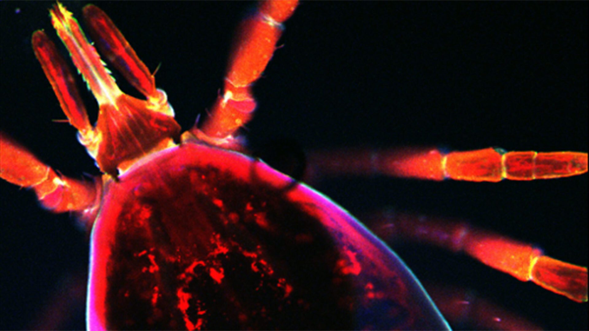
The Borrelia burgdorferi pathogen that causes Lyme disease and the Anaplasma phagocytophilum pathogen that causes Anaplasmosis are two prominent pathogens carried by the black-legged tick, Ixodes scapularis—also known as the deer tick. Dumler is an expert in Anaplasma, while Pal is known around the world for his research with Borrelia. Much remains in question about how both pathogens move from the skin to the blood and then from the blood to their target tissues and organs. Because these processes are profoundly distinct for each organism, it’s nearly impossible to carry out the research effectively in animal models.
The researchers are able to visualize this process in real time, tracking pathogens with fluorescent dyes. They’re able are able to manipulate the models with different types of cells and structures to visualize how transfer of pathogens in and out of the bloodstream occurs.
“Being able to study pathogen dissemination in a configuration that mimics what happens in a real tissue will give a much better picture about what happens in real life,” Dumler said.
